
Plantar Fasciitis, Pathology, Management Tips & Treatment
Pathology
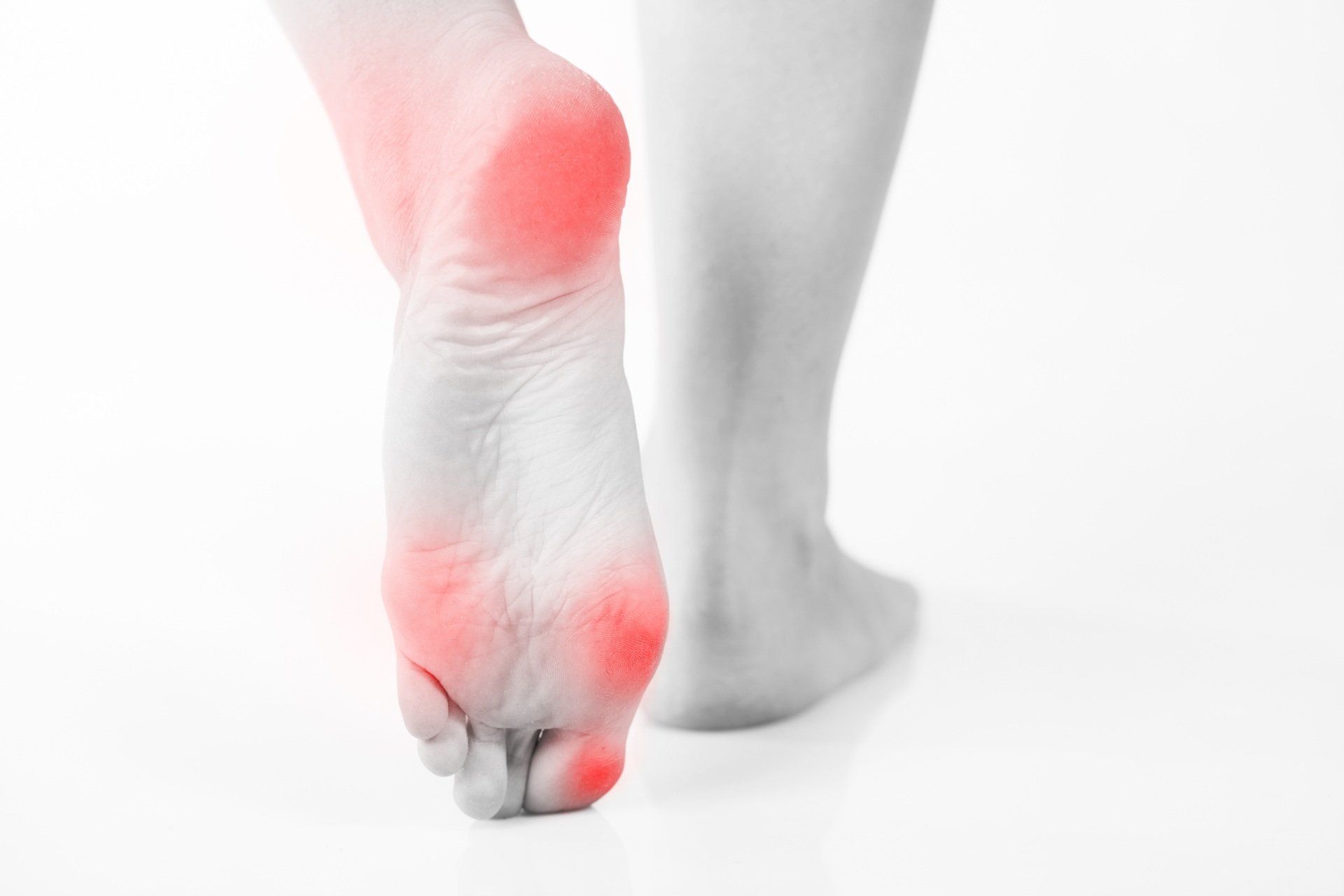
Plantar fasciitis is an overuse condition involving irritation, inflammation, or degeneration of the plantar fascia, the thick band of connective tissue running along the sole of the foot from the calcaneus to the toes. It may affect one or both feet.
Symptoms typically develop gradually, with no specific history of trauma. Pain is commonly worse first thing in the morning or after periods of rest, such as when getting out of bed and may ease with initial activity. Discomfort often increases again with prolonged loading and is frequently most noticeable during the pre-swing phase of the gait cycle.
Pain is usually localised to the plantar aspect of the heel and arch, particularly at the antero-inferior surface of the calcaneus. Associated conditions such as achilles tendinopathy are commonly present due to shared loading patterns within the posterior chain.
Contributing Factors and Causes
Plantar fasciitis is most often linked to excessive or repetitive loading of the plantar fascia. Common contributing factors include:
- Sudden increases in training volume or intensity.
- Poor training technique or inadequate recovery.
- Poor foot biomechanics.
- Weakness or reduced load tolerance in the gastrocnemius and soleus muscles.
- Limited ankle dorsiflexion.
- Inappropriate or worn footwear.
- Increased body weight.
Neural Contributions
In some cases, symptoms resembling plantar fasciitis may be influenced or exacerbated by neural involvement. The sciatic nerve, which originates in the lumbar spine and travels down the posterior aspect of the leg into the foot, can refer pain distally when irritated. Compression due to lumbar disc pathology, spinal stenosis, or piriformis tightness may produce heel or arch pain that mimics plantar fasciitis, often described as burning, sharp, or electric in nature.
Neural tension or irritation can increase sensitivity in the plantar tissues, potentially perpetuating symptoms or reducing the tissue’s ability to tolerate load.
Clinical Presentation
- Localised pain at the anteromedial aspect of the calcaneus.
- Tenderness on palpation of the plantar fascia and possibly the midfoot.
- Palpable thickening or adhesions in chronic cases.
- Pain reproduced with active or passive toe extension due to tension on the plantar fascia and intrinsic foot muscles.
Differential Diagnosis
Not all heel pain is plantar fasciitis. Other conditions that should be considered and ruled out include:
- Fat pad contusion or atrophy
- Calcaneal stress fracture due to repetitive loading or impact
- Tarsal tunnel syndrome, involving entrapment of the posterior tibial nerve at the medial malleolus
Appropriate clinical assessment and referral are essential where indicated.
Treatment Considerations
Client positioning is important. Treating the client in prone allows effective access to compensatory structures in the lower leg. Techniques such as effleurage and petrissage may be applied to hypertonic muscles, particularly the gastrocnemius and soleus.
Muscle energy techniques may be used, including post-isometric relaxation (PIR) or reciprocal inhibition (RI), depending on presentation. In chronic cases, gentle fascial techniques or cross-fibre friction may be applied to areas of adhesion within the plantar fascia.
A global approach is essential. Assessment and treatment should extend up the kinetic chain to include the ankle, lower leg, knees, hips, gluteals, and lumbar spine—particularly in acute or recurrent presentations.
Home Care
Home management is a key component of recovery and should be tailored to the underlying cause. This may include:
- Load management and training modification.
- Home exercises: Stretching or strengthening programmes.
- Footwear assessment or orthotic referral.
- Referral for strength and conditioning support where appropriate.
- Self massage.
Identifying and addressing contributing factors is critical to long-term resolution and prevention of recurrence.


