
Ideology, Anatomy, Pathology & Management
Sciatica, Sciatic Nerve & Anatomy
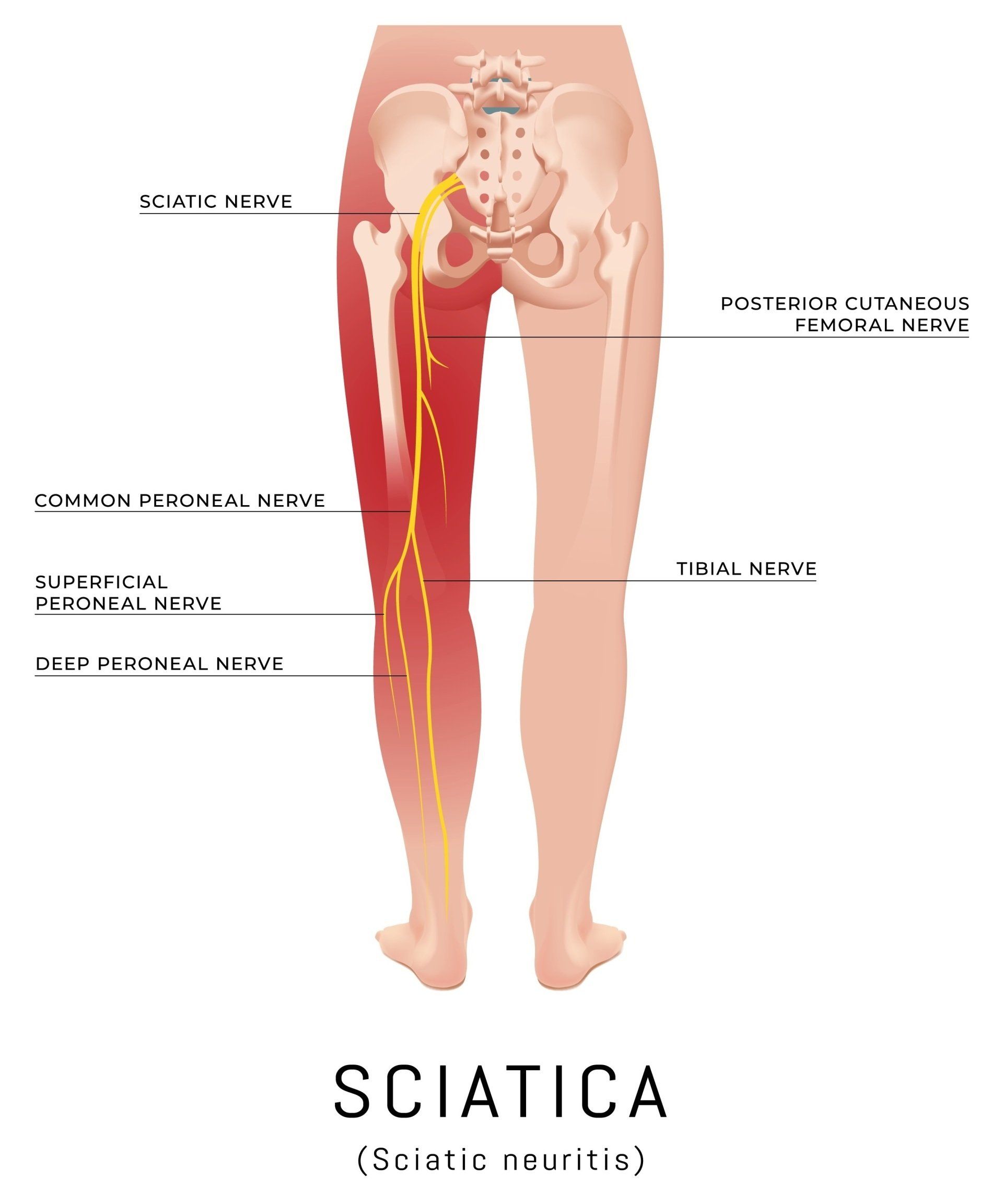
The sciatic nerve (SN) is a Greek work derived from "Ischiadichus" and also known as the ischiadic nerve (18). Renowned for being the longest, widest and largest peripheral nerve in the human body in the medical world, the sciatic nerve supplies most muscles of the leg, skin of the posterior compartment of the thigh, leg and foot. It consists of two components; the tibial nerve (TN) and common peroneal nerve (CPN) component of the sciatic nerve. The union of five spinal nerve roots from the lower lumbar spineand the sacrum form the sciatic nerve and are derived by the joining of the ventral rami L4-S3(18).
Non specific lower back pain (NSLBP) places a substantial economic burden on private and public healthcare providers and direct costs for the provision of care to address NSLBP in the UK has previously been estimated at £11 billion, 51% of which was attributed to costs in primary care, physiotherapy and allied health professions (7).
Sciatica accompanies about 10% of lower back pain episodes and nerve root compression by disc herniation is regarded as the most frequent cause of sciatica (7).It is estimated that 90% of sciatica cases are the result of a disc herniation or disc bulge in the lumbar spine that compresses on the sciatic nerve root (5).
Symptomatic disc herniation is therefore probably a common presentation in osteopathic clinics. Three categories of disc herniation are generally recognised, consisting of disc protrusions (in which the nuclear material protrudes or bulges against the annulus), extrusions (in which the nuclear material escapes the border of the annulus), and sequestration (when the extruded nuclear material loses contact with the remaining disc nucleus)(7).
Sciatica is mainly diagnosed via case history taking and physical examination, individuals may be asked to report the distribution of their pain and whether they are experiencing radiation below the knee. Symptoms of sciatica is commonly a radiating shooting pain and/or the experience of numbness or tingling down the back of one or both legs which is also known as radiculopathy (9, 17).It may include the inability to walk depending upon where the pressure of the sciatic nerve occurs(18).
Statistically, approximately 60% of patients visiting primary healthcare with lower back pain (LBP) report back-related leg pain and it is estimated 5%-10% of patients with LBP have sciatica (9). The annual prevalence of intervertebral disc related sciatica in the general population is estimated to be 2.2% of all cases (9).
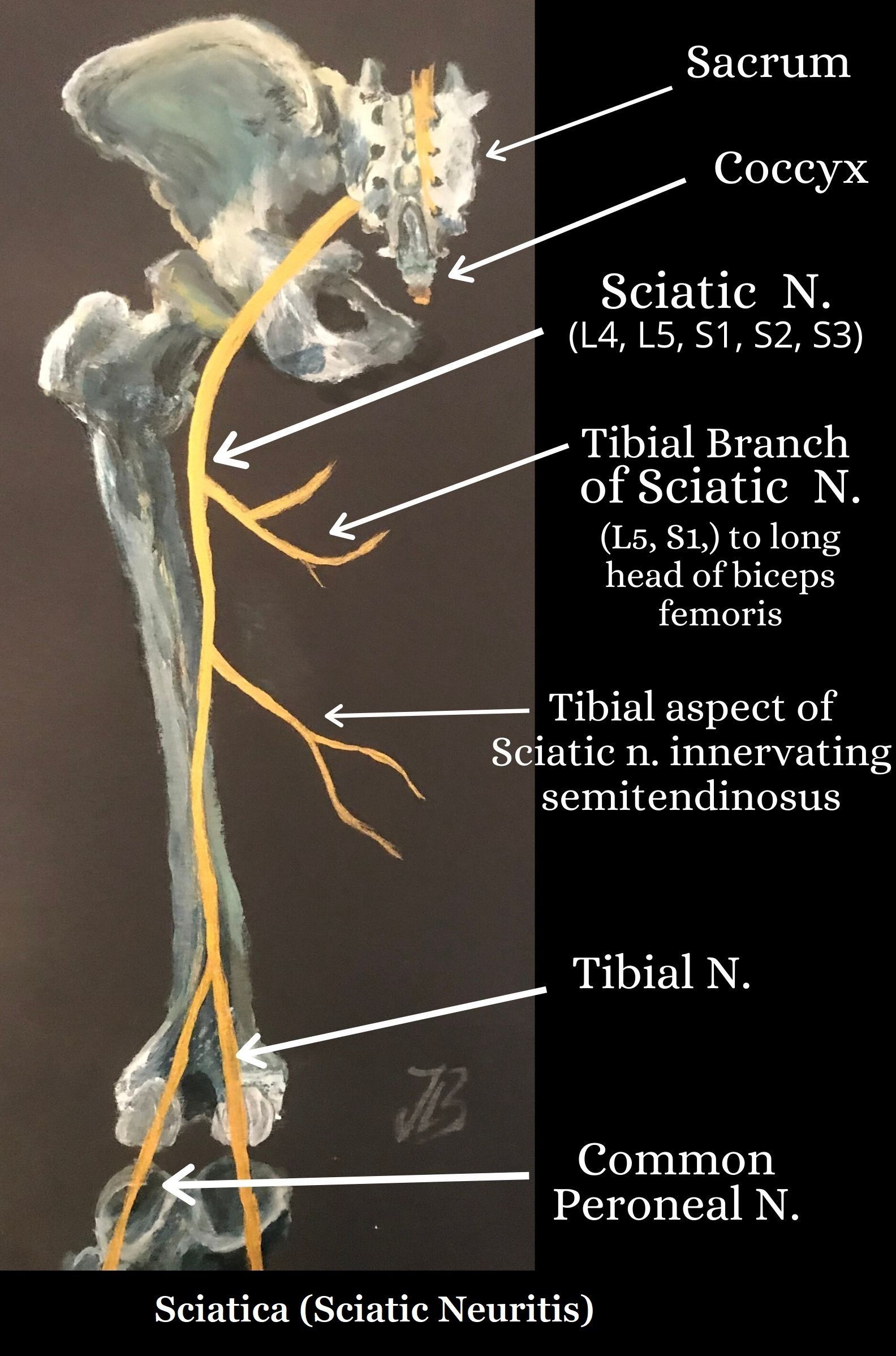
Figure 1. Upper posterior view of the Sciatic Nerve (4, 6)
Risk Factors
- Age: commonly between 45 - 64 years
- Height
- Mental Stress
- Cigarette smoking
- Exposure to vibration from vehicles
- Obesity / being overweight
- Heavy labor / strenous physical activity
- Full time desk jobs
- Occupation factors
- Poor posture (although not proven)
- Strenuous physical activity e.g. frequent lifting, bending and rotating the body.
- Driving, vibration of the whole body.
Smokers have a slower healing rate and recovery process than nonsmokers and have a higher risk of recurrent lumbar disc herniation than nonsmokers(19). Smoking increases the production and release of inflammatory cytokines in the intervertebral discs which interferes with the healing process.
Symptoms
Sciatica is characterised by neuropathic pain radiating from the lower back into the leg that follows the path of the sciatic nerve, often experienced down one or both legs and can sometimes also cause numbness or tingling (paraesthesia). It is often caused when the sciatic nerve (which runs from the back of the pelvis through the buttocks and down to the feet) is irritated, causing pain (16).
- Unilateral leg pain is greater than lower back pain (9).
- Radiated pain below the knee into the foot or toes with either numbness or paraesthesia in the same distribution.
- Referred leg pain not related to a nerve root pathology (10).
- Radiated pain with a dermatomal pattern.
- Lower back pain.
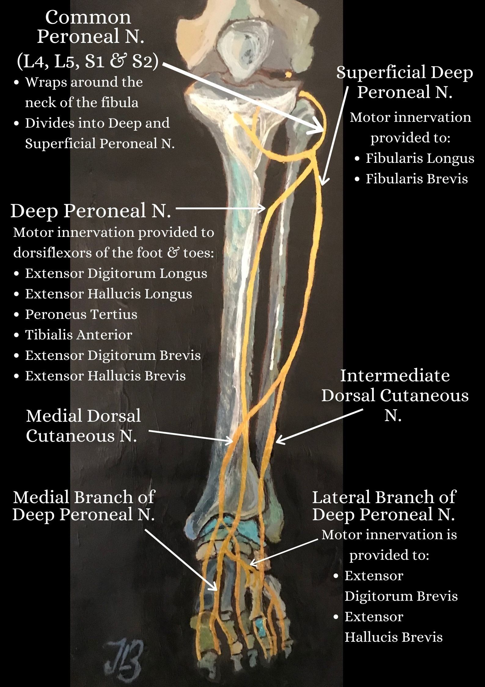
Figure 2. Lower anterior view of the Sciatic Nerve (4, 14, 16)
Causes
- Herniated Intervertebral Disc: Herniated lumbar disc is a displacement of disc material (nucleus pulposus or annulus fibrosis) beyond the intervertebral disc space (8).
- Sciatic Nerve Entrapment: May occur from the pelvis to the distal thigh. The most common site is between the greater sciatic notch and ischial tuberosity and by the piriformis muscle. Myositis ossificans (bone tissue formation inside muscle) of the biceps femoris muscle is another possible site. Biceps femoris muscle insertion on the linea aspera to the adductor magnus muscle is also another possible entrapment site to compress the sciatic nerve (14).
- Piriformis Syndrome: The entrapment of sciatic nerve as it exits the greater sciatic notch islikely due to a myospasm or contracture of muscles i.e. piriformis or gemellus superior muscle (4).
- Lumbar Spinal Stenosis (LSS): The anatomical narrowing of the spinal canal which can be associated with various clinical signs and symptoms including back pain and claudication (19). Patients frequently complain of numbness and pain within the lumbar region and lower limbs along with intermittent neurogenic claudication caused by compression of the nerve roots with narrowing of the spinal canal and the intervertebral foramen which can lead to gait disorders (11).
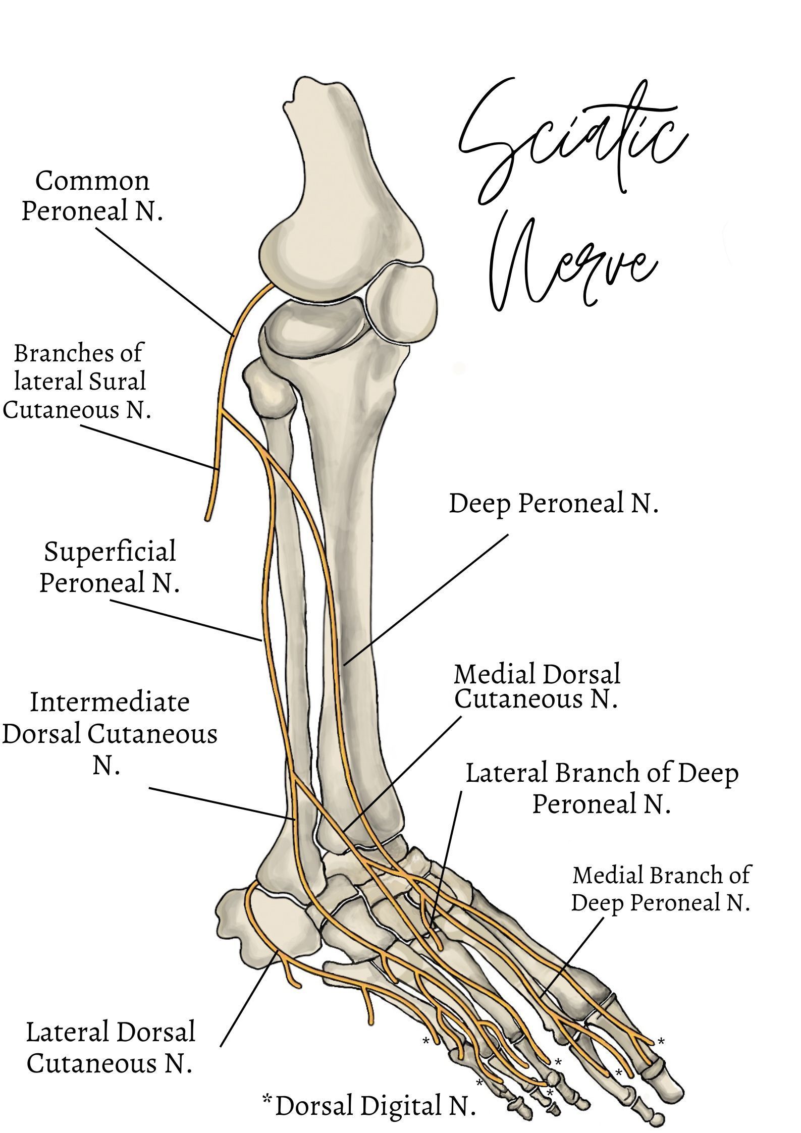
Figure 3. Lower lateral view of the Sciatic Nerve (15, 21)
Neural Course, Cutaneous & Motor Innervation of the Sciatic Nerve
The sciatic nerve (SN) is formed in the pelvis by the joining the ventral rami L4-S3 SN roots (22) (figure 1). It travels inferiorly into the pelvic surface of the coccygeus muscle and enters the gluteal region. The SN leaves the pelvis through the greater sciatic foramen below the piriformis muscle and passes deep to the gluteus maximus
muscle before descending between the greater trochanter and ischial tuberosity in the gluteal region. The trunk of the sciatic nerve innervates the proximal parts of all three hamstrings muscles (semi membranosus, Semi tendonosus and Biceps Femoris) and ischial aspect of the adductor muscles (4).
The sciatic nerve then divides at the apex of the popliteal fossa into two main branches; the tibial nerve
and the common fibular nerve
(figure 1).
After the SN bifurcation, the tibial component innervates the semi membranosus, semi tendinosus, long head of biceps femoris muscles and adductor magnus. The short head of the biceps femoris is innervated via the common peroneal component of the SN.
Common
Peroneal Nerve
The common fibula (peroneal) nerve contains posterior fibres L4 and L5 of the lumbosacral trunk and lumbar sacral roots S1 and S2. It then crosses over plantaris and the lateral head of the gastrocnemius to gain access of the anterior leg before wrapping around the neck of the fibula and passing through the peroneus longus, where it bifurcates and divides into two terminal branches;
the superficial
and deep fibula
(peroneal) nerve
(figure 2 and 3)(4).
Superficial Peroneal Nerve
Provides motor innervation to the following muscles (4):
- Peroneus longus
- Peroneus brevis
- Cutaneous (skin) supply to the dorsum of the foot.
- Cutaneous (skin) inferior 1/3 of the leg on its medial and lateral aspects.
Deep Peroneal Nerve
Travels inferolaterally towards the interosseus membrane and innervates the muscles below (4):
- Extensor digitorum longus
- Extensor hallucis longus
- Fibularis tertius
- Tibialis anterior
- Extensor digitorum brevis
- Cutaneous innervation to the first interdigital cleft of the foot.
- Cutaneous sensation between the first and second toes.
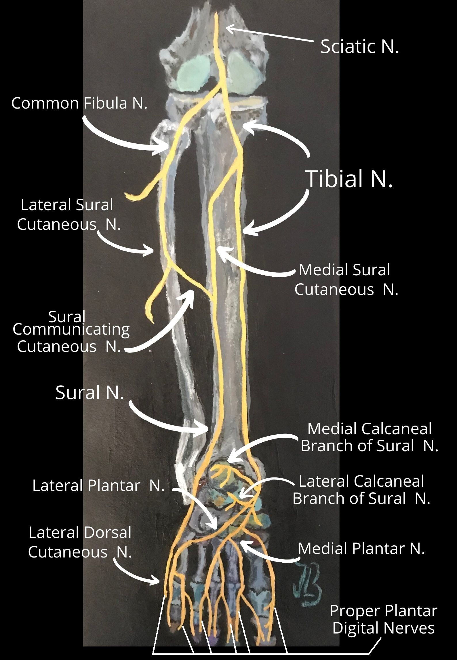
Figure 4. Lower posterior view of the Sciatic Nerve (4, 6, 15)
Tibial Nerve
The tibial nerve is known as the medial terminal branch of the sciatic nerve and contains anterior nerve fibres of the ventral rami L4 and L5 from the lumbosacral trunk and the first three sacral nerves S1, S2 and S3 (4).
The tibial nerve crosses medially over the tibial vein to rest between the tibial vein and tibial artery on the superficial surface of the popliteus muscle and enters the leg under the tendon of the soleus. After the bifurcation of the sciatic nerve at the apex of the popliteal fossa, the tibial nerve travels inferiorly straight through the fossa and gives off muscular branches to the following muscles:
- Soleus
- Plantaris
- Popliteus
- Gastrocnemius (medial and lateral heads)
- Tibialis posterior
- Flexor Hallucis longus
- Flexor Digitorum longus
- superior tibiofibular joint, tibia bone, interosseous membrane of leg, and the inferior tibiofibular joint.
As the tibial nerve travels inferiorly accompanied by the tibial vessels, it pierces the tendinous arch of the soleus to continues between and innervate the muscles:
· Flexor digitorum longus
· Tibialis posterior
· Flexor hallucis longus
- From the middle of the popliteal fossa, the medial sural cutaneous nerve branches from the tibial nerve and travels between the two heads of the gastrocnemius muscle before descending inferiorly to the posterior lateral aspect of the malleolus and terminate at the lateral 5th metatarsal bone (4).
- Muscles supplied: triceps surae, plantaris, popliteus, tibialis posterior, flexor digitorum longus and flexor hallucis longus muscles.
The medial and lateral sural nerves are made up of collateral branches from both the tibial and common peroneal nerves and provide sensation to the calf and a small lateral portion of the foot (4, 7).
Conditions Related To An Injured Sciatic Nerve
Tarsal Tunnel Syndrome
Compression of the tibial nerve or either of its terminal branches (lateral or medial plantar nerve)in the tarsal tunnel leads to a condition known as tarsal tunnel syndrome. A patient with the syndrome may experience plantar pain and sensory disturbances and can also result in palsies of the intrinsic foot muscles (4).
Injury to the Deep Fibular Nerve
An isolated lesion of the deep fibular nerve can lead to muscular innervation being affected and cause the prevention of dorsiflexion resulting in foot drop and a steppage gait (4).
A steppage gait is a gait abnormality that is often adopted to counteract the effects of foot drop by exaggerating the flexion at the hip and knee during walking to allow the toes to clear the ground.
The tibial nerve then passes through the tarsal tunnel of the medial ankle and divides into two terminal branches; the medial plantar nerve and the lateral plantar nerve. These two terminal branches of the tibial nerve supply all the muscles on the plantar aspect of the foot (4).
Injury to the Common Fibular Nerve
The sciatic nerve is the most commonly injured nerve in the lower limb and because of its subcutaneous position around the head of the fibula, it is vulnerable to direct trauma (17, 4).
Damage to the common fibular nerve at the neck of the fibula can cause weakness or paralysis of the muscles in the anterior and lateral compartments of the lower limb. This can cause a loss of dorsiflexion which often results in foot drop.
Foot drop
Foot drop is a type of gait abnormality in which the toes cannot clear the ground during walking. If damage to the common fibular nerve occurs after it divides into its terminal branches, symptoms are dependent at which the terminal branches have been affected. For example, an isolated lesion of the superficial fibular nerve generally affects sensory innervation which can cause pain in the distal leg and the dorsum of the foot (4).
Video 1. Foot drop presentation. We are very grateful and thank the individual's consent to display their walking gait.
Diagnsosis & Imaging
Diagnostic imaging such as an MRI is only useful for if the results influence an individual’s further management. It may also be required for if patients fail respond to conservative care (i.e. physical therapy) for 6 to 8 weeks (9).
The straight leg raise (SLR) is one of the most commonly performed physical tests during clinical examinations to test for sensitivity and impairment of mechanical function of the sciatic nerve and the lumbosacral nerve roots in individuals with low back or lower extremity pain (3). The test is designed to apply tension to the sciatic nerve and lumbosacral nerve roots to evoke a physiological response such as reported pain, neural involvement and/ or altered sensations(3).
Treatment
The primary aim of conservative treatment for sciatica from a physical therapist is to reduce the pain and reduce the pressure on the sciatic nerve root. Initially, intervention for the treatment of patients with sciatica is a nonoperative approach which has demonstrated to be beneficial in more than 50% of patients (13).
Corticosteroid Injections
Epidural corticosteroids are administered locally
at the site of a lesion are likely to be effective during the
first weeks of an episode of sciatica.Compared with no epidural corticosteroid, epidural corticosteroids may be more effective at improving limb pain at
2 weeks, but may be no more effective after more than 2 weeks in people with disc herniation (8).
Surgery
Surgical intervention is often considered for if symptoms persist after 8 to 12 weeks and is generally regarded as the final or "last resort" in what is in many cases a very long journey through failed medical management (13). Surgical intervention for sciatica focuses on removing the disc herniation via the removal of part of the disc or foraminal stenosis and is the choice for unilateral sciatica. The aim of the treatment modality is to ease the leg pain, but the predominant focus is removing the lower back pain (9). Cauda equina syndrome is an indication for the need of immediate surgery.
Surgical intervention has been compared to conservative treatment for patients with sciatica. Koe et al.,
(2009) found that surgical intervention may provide a quicker relief of symptoms after one year and gain quicker relief of leg symptoms than patients receiving conservative treatment. But, no significant differences were found in terms of success rate in one or two years time during the patients' follow up, in some cases after 4 and 10 years follow up (9).
From a therapist's perspective, the success rate and outcome may vary from one individual to another and factors that need to be considered is the patient's severity of their present condition, lifestyle, previous exercise history and whether they have any underlying conditions which may or may not have been diagnosed whilst having consulted a practitioner. Patient preference for treatment modalities and methods will also differ.
Both standard discectomy
and microdiscectomy
seem to increase self-reported improvement to a similar extent(8).
Conservative Management
Additional options include careful use of anti-inflammatory medications, physiotherapy, massage therapy, local injections, acupuncture and chiropractic treatment. There is no clear medical or scientific evidence as to whether particular interventions do help sciatica or lumbar disc herniation. From my clinical experiene, patient education is of paramount importance with encouraging clients to understand how to manage their injury. Recovery rates are unique and individual for everybody as lifestyle factors, exercise history, body biomechanics and posture all play pinnacle roles.
For example, a 47 year old gentleman, Bill sits working at a computer desktop for 9 hours, 5 days a week. His diet consists mainly of convenience foods high in processed fats and salts, performs less than 2 hours of weekly exercise by walking to work from his car and back at the end of a long day. His recovery rate is predicted to be slower than 29 year old Andrew who is a full time postman, has an active lifestyle, has healthy diet and has attended a gym four times a week for the past ten years and plays a sport.
Spinal Manipulation
Spinal manipulation for sciatica has been found to help improve patient outcomes, some studies have found by up to 50%(13).
How Could Osteopathy Help You?
Osteopaths are providers of manual therapy, exercise, self-management and lifestyle advice, which are interventions recommended in the most recent NICE Non-Specific Low Back Pain (NSLBP) and sciatica guidelines (7). Practitioners are frequently consulted for management of spinal symptoms, with NSLBP; the most common symptom presented to osteopaths in the UK (7).
There is debate regarding the most effective treatment of NSLBP as no one treatment modality is considered to demonstrate significant benefits over others. However, treatment following NSLBP guide-line recommendations has shown clinical and economical benefit compared with non-standardised treatment (7).
Disc herniation has frequently been thought of as a surgical disorder, but opinion in much of the literature is that conservative care should be tried initially, in the absence of indications for immediate surgery such as cauda equina syndrome or rapid and progressive neurological deficits (7). A variety of conservative measures are used including oral medication, epidural injections, bed rest, traction, electrotherapy, exercises, back school or courses, acupuncture and manipulation (7). Many of these approaches are of uncertain efficacy, and manipulation is no exception. Even surgical approaches such as microdiscectomy and laser discectomy have not been shown to be more effective than standard care (7).
References
- Adibatti, M., V. S. (2014) Study on Variant Anatomy of Sciatic Nerve, Journal of Clinical and Diagnostic Research, 8; 8: 7-9.
- Anbumani, T. L., Thamarai Selvi, A., Anthony Ammal S. (2015) Sciatic nerve and its variations: an anatomical study. Int J Anat Res.;3 (2): 1121–1127.
- Bueno-Gracia, E., Estébanez-de-Miguel, E., López-de-Celis, C., Shacklock, M., Caudevilla-Polo, S., González-Rueda, V., Pérez-Bellmunt, A. (2020) Effect of ankle dorsiflexion on displacement and strain in the tibial nerveand biceps femoris muscle at the posterior knee during the straight leg raise:Investigation of specificity of nerve movement,
- Crumbie, L. (2019) [online] Sciatic Nerve: Kenhub.com , last viewed 31/05/2020; www.kenhub.com/en/start/anatomy-sciaticnerve-branches.
- Frost, L. R., Brown, S. H. M. (2016) Muscle Activation Timing and Balance Response in Chronic Lower Back Pain Patients with Associated Radiculopathy, 32; 124-130.
- Gilroy, A. M., Macpherson, B. R., Ross, L. M. (2008) Atlas of Anatomy, Thieme, New York.
- Inman, J., Thomson, J. P. (2019) Complementing or Conflicting? A Qualitative Study of Osteopaths’ Perceptions of NICE Low Back Pain and Sciatica Guidelines in the UK, IJOM, 31: 7–148.
- Jordan, J., Konstantinou, K., O'Dowd, J. (2009) Herniated Lumbar Disc, Clinical Evidence, 03; 1118: 1-33.
- Koes, B. W., Tulder, M. W., Peul, W.C. (2007) Diagnosis and Treatment of Sciatica; Clinical Review, 334: 1313-1317.
- Konstantinou, K., Dunn, K. M., Ogollah, R., Lewis, M., Windt, D. v. d., Hay, E. M. (2018) Prognosis of Sciatica and Back-Related Leg Pain in Primary Care: the ATLAS Cohort, The Spine Journal, 18: 1030-1040.
- Kuwahara, W., Deie, M., Fujita, N., Tanaka, N., Nakanishi, K., Sunagawa, T., Asaeda, M., Nakamura, H., Kono, Y., Ochi, M. (2016) Characteristics of Thoracic and LUmbar Movements During Gait in Lumbar Spine Stenosis Patients Before and After Decompression Surgery, Clinical Biomechanics, 5: 15-19.
- Lewis, S., Jurak, J., Lee, C., Lewis, R., Gest, T. (2016) Anatomical variations of the sciatic nerve, in relation to the piriformis muscle, Translational Research in Anatomy,
- McMorland, G., Suter, E., Casha, S., du Plessis, S.,Hurlbert, R. J. (2010) Manipulation or Microdiskectomy for Sciatica? A Prospective Ranomised Clinical Study, Journal of Manipulative and Physiological Therapeutics, 33; 8: 576-584.
- Monteleone G, Stevanato G. Entrapment of the sciatic nerve at the linea aspera: A case report and literature review. Surg Neurol Int 19-Oct-2016;7:89. Available from: https://surgicalneurologyint.com/surgicalint-articles/entrapment-of-the-sciatic-nerve-at-the-linea-aspera-a-case-report-and-literature-review/
- Netter, F. H. (2014) Atlas of Human Anatomy: 6th Ed., Elseiver Saunders, USA.
- NICE guideline (2016) [ONLINE] Low back pain and sciatica in over 16s: assessment and management, https://www.nice.org.uk/guidance/ng59/ifp/chapter/What-are-low-back-pain-and-sciatica, last viewed 28/05/2020.
- Palastanga, N. P., Field, D., Soames. R. (2006) Anatomy and Human Movement: Structure and Function [5th Ed.], Elsevier.
- Saritha, S., Praveen Kumar, M., Supriya, G. (2012) Anatomical Variations in the Bifurcation of the Sciatic Nerve, A Cadaveric Study and its Clinical Implications, Anatomy & Physiology: Current Research, 2; 5: 1-4.
- Shiri, R., Falah-Hassani, K. (2016) The Effect of Smoking on the Risk of Sciatica: A Meta-Analysis, The American Journal of Medicine, 129: 64-73.
- Siebert, E., Prüss, H., Klingebiel, R., Failli, V., Einhäupl, K. M., Schwab. J. M. (2009) Lumbar Spinal Stenosis: Syndrome, Diagnostics and Treatment, Nature Reviews; Neurology, 5: 392-403.
- Toms, A. F., Rushton, L. A., Kennedy, N. R. (2017) Muscle Herniation of the Peroneus Longus Muscle Triggering Superficial Fibular Nerve Paresthesia, Sonography, 5: 1. Wiley Online Library; www.onlinelibrary.wiley.com
-
V, S., Adibatti, M. (2014) Study on Variant Anatomy of Sciatic Nerve,Journal of Clinical and Diagnostic Research, 8; 8: 7-9.


